Reading is one of the most complex cognitive tasks the human brain performs. From recognising letters as symbols, constructing meaning, engaging emotions, to managing attention and memory—reading requires the synchronous operation of many brain regions. But what happens when this delicate harmony is disrupted?
Thanks to qEEG and Neuromap® analysis, we can now observe, analyse, and understand both typical and atypical reading functions with unprecedented accuracy.
In this article, we’ll share a key finding from our team’s recent work that reveals how children with reading difficulties show unique patterns of brain connectivity. We’ll explore how Neuromap identifies the neurological roots of reading delays and disorders—and how this insight is transforming education, therapy, and personalised interventions.
Understanding Reading in the Brain
Reading is not housed in a single region of the brain. It emerges from the coordination of several key areas:
Occipital lobes: responsible for visual processing
Temporal lobes: involved in auditory perception and phonological decoding
Parietal lobes: contribute to spatial awareness and multisensory integration
Frontal lobes: manage working memory, attention, planning, and executive functions
For reading to be effective, these regions must communicate with each other in a balanced and flexible way. This is where connectivity comes into play.
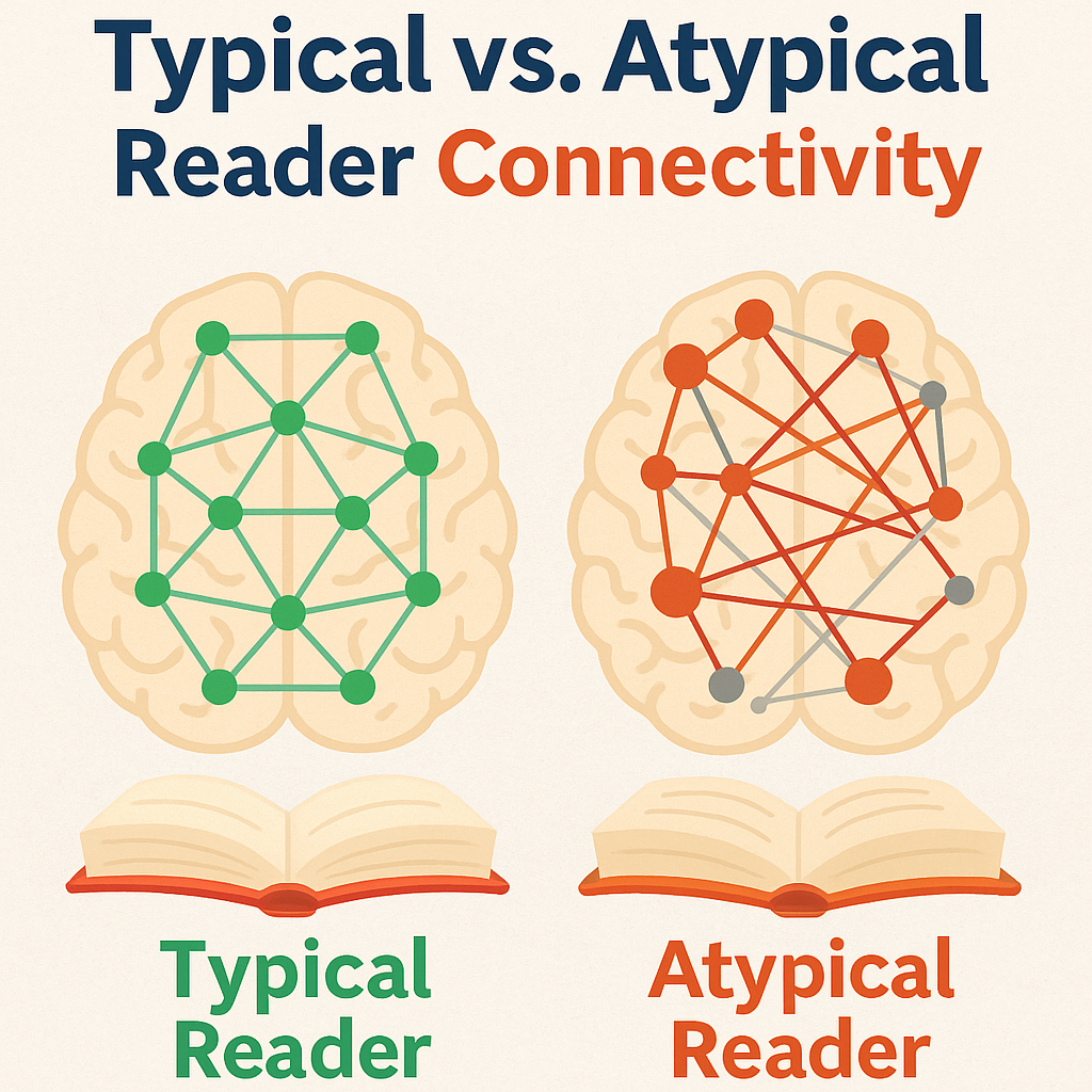
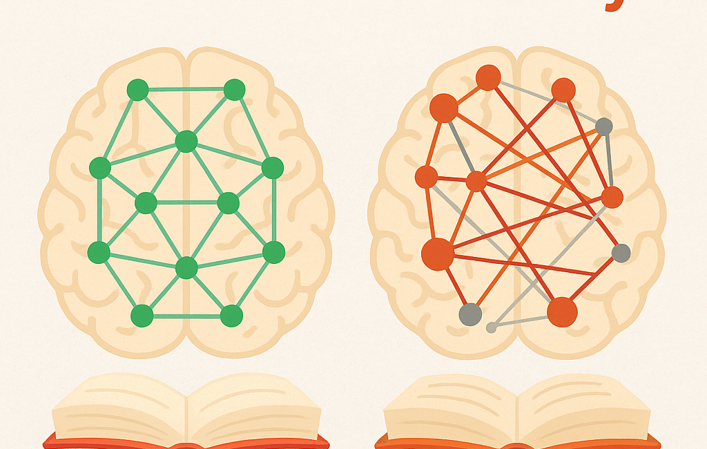
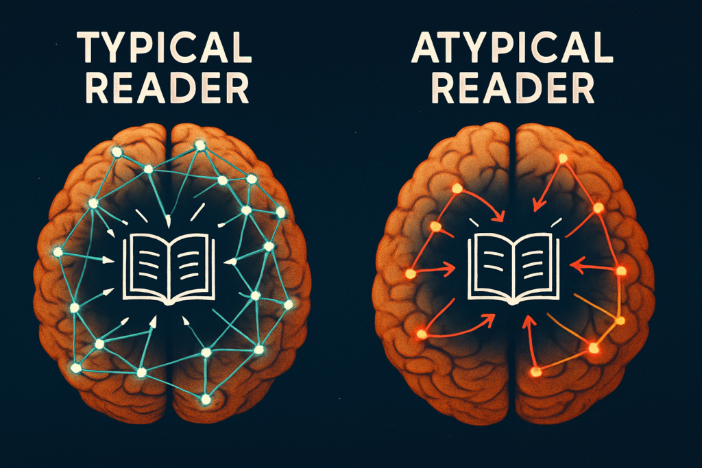
Typical vs Atypical Reading: A Connectivity-Based Comparison
Reading may appear simple, but it is an intricately orchestrated process involving simultaneous, coordinated activity across multiple brain regions. To recognise letters, match them to sounds, build words, and understand meaning, the visual, auditory, linguistic, and cognitive areas of the brain must maintain well-balanced communication. We call this brain connectivity.
Brain Connectivity in Typical Readers
In children with healthy reading skills, we often observe:
Moderate coherence between the left temporal (T3) and parietal (P3) lobes. This supports the child’s ability to decode the sounds and meanings of words. When they see the letter “k”, they instantly recognise its sound and place it correctly in the word “cat”.
Strong bidirectional communication between the F7 region and Broca’s area (BA 44/45). These regions are responsible for speech and language production. This connection allows the child to read aloud fluently and understand sentence structure.
Dynamic coordination between Cz (central hub) and the visual cortex (O1, O2). This controls eye tracking across lines of text and helps the child move smoothly from one word or sentence to the next.
Such children don’t just see text—they process it, internalise it, and can recall it when needed.
Brain Connectivity Issues in Atypical Readers
Children with reading difficulties show either insufficient or excessive connectivity. Let’s break this down:
Low connectivity between temporal and parietal regions (hypocoherence)
This can impair a child’s ability to break words into individual sounds or match letters to sounds. They may confuse letters like “b” and “d”, or struggle to syllabify words like “butterfly”.
Parents might notice their child repeatedly forgetting a word or reading the same word as if it were new each time.Excessive connectivity in the frontal lobes (hypercoherence)
This suggests the brain’s executive areas are overly engaged and locked into repetitive loops. These children may get “stuck” on a word or line, reading it over and over, unable to move on.
Parents often say, “It’s like his brain freezes,” or report emotional overwhelm and anxiety during reading.Insufficient connectivity between visual and language regions
Low coherence between the visual cortex (O1, O2) and language-processing areas (such as F7 and T3) can make it difficult to recognise letters and form words. Letter reversals, slow decoding, and frequent mistakes are common.
For example, a child might read “The dog ran” as “he god ran,” mixing up sounds and order due to poor visual-language integration.
A Practical Observation Guide for Families
| Symptom | Connectivity Issue | What Parents See at Home |
| Confusing letters, forgetting sounds | Weak connection between temporal-parietal | Skipping or misreading letters or syllables |
| Reading the same line repeatedly | Hyperconnected frontal regions | Getting stuck, struggling to move between tasks |
| Reversing letters or slow word recall | Low visual-language integration | Saying “b” for “d”, slow or hesitant reading |
| Avoiding reading, low confidence | Imbalance in frontal/temporal networks | “I can’t do this,” or refusing to read aloud |
Important Note for Parents: These behaviours are not due to laziness, defiance, or lack of interest. When the brain’s communication systems are either too weak or too strong, the child cannot control these difficulties consciously. Neuromap helps us understand what’s happening beneath the surface—offering a clear window into a child’s unique brain.
Every brain is different. So is its connectivity. Through tailored solutions based on each child’s brain profile, we can make the reading journey easier, more efficient, and even enjoyable.
Neuromap Analysis: Making the Invisible Visible
Neuromap® uses qEEG data to create real-time maps of brain connectivity. It pinpoints which areas are out of sync, which are overactive, and which are underdeveloped. Based on this, we can design targeted interventions using methods like:
tDCS (transcranial direct current stimulation)
Neurofeedback
Sensory-motor integration therapy
One consistently observed pattern is the disconnection between T5 (left posterior temporal) and P3 (left parietal), which often correlates with dyslexia-like symptoms. Another is low synchrony between Cz and O1, which reflects difficulties in visual tracking and scanning.
What This Means in the Classroom
In many traditional classrooms, reading struggles are misunderstood as laziness, distraction, or behavioural problems. Neuromap helps us go beyond assumptions and see the neurological truth.
Examples:
A student who avoids reading aloud may not be shy—they may have a weak connection between T3 and F7, making it difficult to process sounds.
A child who rereads the same line might not be inattentive—they may have a coordination problem between the frontal lobe (Fz) and the occipital cortex (O1, O2), affecting visual flow and comprehension.
By identifying these patterns, teachers and therapists can personalise reading instruction to the child’s neurological needs.
Why Neuromap Is a Game-Changer
Evidence-Based Decisions
Educators and therapists no longer have to rely on guesswork—they can use data to guide interventions.Early Identification
Children at risk of falling behind can be identified before they struggle academically.Tailored Strategies
Every child’s reading circuit is unique. Interventions must reflect this individuality to be truly effective.
The Bigger Picture: Not Just Reading, But All Learning
Reading is just one cognitive skill we can measure. Neuromap’s power lies in its ability to assess multiple domains:
Language development
Attention and focus
Emotional regulation
Memory and processing speed
By viewing the brain as an integrated system, we stop addressing surface-level symptoms and begin resolving the core problems.
This article reflects just one breakthrough in our work. Every child’s brain reveals new insights. Our mission is to bring those insights to every family, teacher, and therapist. With Neuromap, we no longer guess—we understand. We don’t just observe challenges—we solve them.
Conclusion
With advanced neurotechnologies like Neuromap®, we are entering a new era in neuroeducation. Children with reading difficulties are no longer hidden behind labels. We now have the tools to look into the brain, understand its language, and support its growth in ways that are precise and compassionate.
And this… is just the beginning.
This article is part of our ongoing efforts to merge neuroscience with education. All data is anonymised and shared solely for educational and clinical awareness purposes.