The Neuromap® connectivity analysis offers a profound transformation in how we understand and approach the brain, especially in children with autism and other neurodevelopmental conditions. Rather than simply viewing brain regions in isolation, Neuromap® reveals how these areas connect and communicate,or fail to. Imagine the brain as a vast orchestra, with different sections playing their parts. Some instruments might be silent, others might be playing too loudly, and some may be out of sync altogether. Neuromap® captures this intricate performance by analyzing coherence,the harmony or dissonance between signals from different parts of the brain. This coherence is not just a technical term; it represents how well two brain regions are working together, and this has a direct impact on how a child feels, learns, communicates, and responds to their world.
Disconnection between brain regions, also referred to as hypocoherence, can occur anywhere in the brain. It could be in the frontal lobes, which guide planning and emotional regulation. It could be in the temporal lobes, responsible for processing language and memory. It might occur in the sensory integration regions at the back of the brain, which help children process touch, movement, sound, and visual input. When two areas that should be exchanging information are not doing so, it’s like a phone line being cut. One region might be trying to send a message, but the other never receives it. For a child, this might manifest as emotional detachment, a flat affect, lack of appropriate reactions to social cues, or struggles in organizing their thoughts or actions.
But just as some regions may not be communicating enough, others might be communicating too much. This is what Neuromap® identifies as hypercoherence,an overconnection between two or more areas that become locked in a repetitive loop. This is commonly seen in children who exhibit rigid behaviors, obsessive thoughts, or heightened anxiety. For example, when a child is caught in repetitive hand flapping, what we are often witnessing is the result of sensory and motor regions being excessively synchronized. These parts of the brain are acting like a feedback loop that cannot be turned off. It’s not a behavioral problem in the traditional sense,it’s a biological rhythm, an over-connection that has hijacked flexibility and spontaneity.
The beauty of the Neuromap® model lies in its ability to detect these patterns across multiple brainwave frequencies. It does not offer a single snapshot, but a dynamic, multi-dimensional view of the brain’s communication landscape. Each frequency,delta, theta, alpha, beta, and gamma,reflects different cognitive and emotional processes, and disruptions in these frequencies help pinpoint where communication is failing or stuck. A child might have too little theta between regions involved in memory and focus, or too much beta between areas responsible for controlling fear, resulting in excessive worry or reactivity. These variations are never one-size-fits-all, and they change depending on the child’s developmental stage, emotional environment, and even their sensory sensitivities.
For parents, it’s important to understand that Neuromap® does not label your child with a fixed diagnosis. Instead, it provides a real-time map of their brain’s functional pathways, highlighting where help is needed and where strength already exists. Disconnections are not failures. They are opportunities,clues that tell us where the brain needs reconnection and balance. When these areas are identified, targeted neuromodulation techniques like transcranial direct current stimulation (tDCS) or neurofeedback can be used to gently encourage healthier rhythms. Over time, as these interventions help regions synchronize more effectively, children may show improvements in language, self-regulation, social interaction, and flexibility of thought and behavior.
The changes are often subtle at first, like a softening of gaze, a pause before a repetitive action, or an unexpected attempt at eye contact. But over weeks and months, families begin to witness profound shifts. A child who once screamed when music played may begin to hum along. One who could not tolerate light touch might start to seek hugs. These are not simply milestones,they are reflections of deeper neurological healing, evidence that disconnection is being replaced with meaningful connection. Families are no longer left to guess whether a therapy is working. The Neuromap® allows clinicians to measure change, adjust strategies, and show progress,not only in behavior but in the brain itself.
What makes Neuromap® truly empowering is that it doesn’t just offer data,it offers understanding. It helps parents move away from seeing behaviors as problems and toward viewing them as expressions of the brain’s internal wiring. It encourages hope, not just by promising results, but by honoring the uniqueness of each child’s neurological identity. It tells families: your child is not broken. Their brain may be wired differently, but with the right support, they can find their rhythm, their voice, their balance. And in doing so, they can begin to write a new story,one grounded in their own potential.
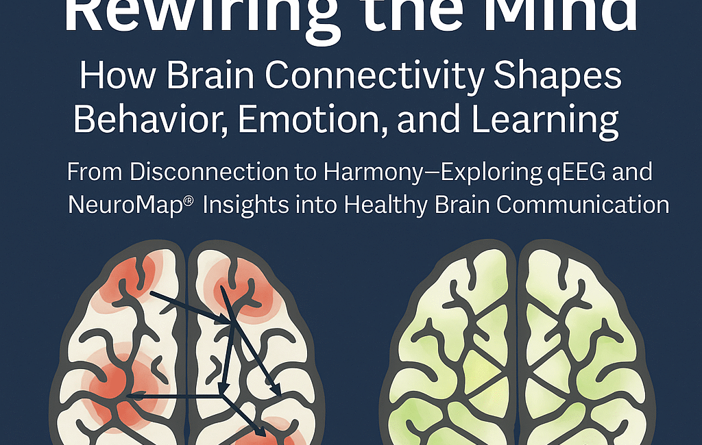
From Disconnection to Harmony, Exploring qEEG and Neuromap® Insights into Healthy Brain Communication
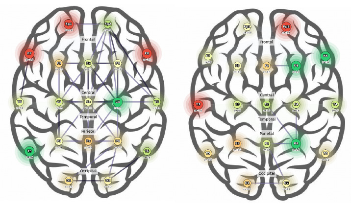
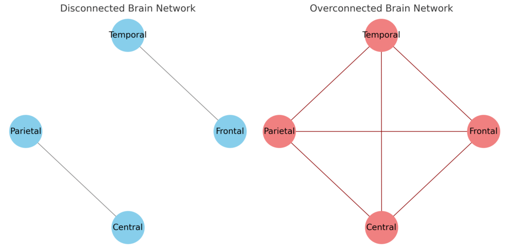
When we compare two different Neuromap® connectivity profiles,one with widespread disconnection across brain regions and another with increased connectivity,we begin to understand not only how the brain struggles, but also how it can begin to heal.
In a highly disconnected brain, such as in the case of Patient Example 1, we observe a pattern where multiple regions across the frontal, temporal, parietal, and central lobes are not properly communicating. These “cut lines” mean that vital pathways,like those that help us plan, speak, understand, and respond,are simply inactive or inefficient. For example, this individual shows disconnections in areas such as Fp1, Fp2, F7, and F3 within the frontal lobe. These areas are crucial for decision-making, language production, and emotional control. A person with these disconnects might struggle to maintain focus, form sentences, or regulate impulsive behavior. It might feel like driving a car with no brakes and no GPS,the brain can move, but it lacks control and direction.
In contrast, in a brain where connectivity is increased but poorly balanced,as seen in Patient Example 2,we observe a different kind of struggle. The frontal regions, including Fz, F4, and Fp2, are not disconnected but overly synchronized. These regions are “talking” so much that flexibility and adaptability are lost. This may lead to anxiety, repetitive thoughts, or an inability to move fluidly from one task or emotion to another. The parietal and occipital regions in this brain also show signs of over-communication, which could lead to visual processing overload or misinterpretation of sensory information. Imagine trying to function when your brain feels like a room with all the lights turned on,bright, intense, and overwhelming.
In a disconnected state, a person might not be able to initiate or maintain meaningful communication. They may experience blank moments during conversations, have difficulty recalling verbal information, or even appear disengaged in their environment. For example, disconnection in the left temporal lobe (T3 or T5) can manifest as trouble understanding spoken language, while disconnection in the right parietal area (P4) can impair spatial awareness or lead to clumsiness when navigating space. A disconnection in Cz or C3 may cause problems with motor coordination,simple tasks like tying shoes or writing can feel disjointed.
On the other hand, a brain with increased coherence can often perform structured, repetitive tasks well,like data entry or detailed art,but may struggle with anything that demands shifting attention or spontaneous interaction. For instance, Patient Example 2’s hyperconnectivity between Fp2 and F4, as well as Fz and C4, means they may become stuck in certain cognitive patterns. They may ruminate on intrusive thoughts or find it difficult to let go of negative emotions, reinforcing cycles of anxiety or pessimism. These over-synchronized circuits can make emotional regulation feel like trying to stop a train that has no brakes.
Where disconnection often results in under-functioning,silence, inaction, or confusion,overconnectivity leans toward over-functioning, hypervigilance, anxiety, or overreactions. Neither is optimal, but both provide valuable insight. The goal of Neuromap® and the QPAN® model is not to force the brain into a predefined mold, but to rebalance it,to strengthen weak pathways, quiet down the noisy ones, and help the brain find its natural rhythm again.
In a more connected and regulated brain, someone like Patient Example 1 could begin initiating speech, understanding verbal instructions, and navigating social settings with increased confidence. Their ability to regulate impulses would improve, leading to fewer hyperactive episodes. Similarly, for someone like Patient Example 2, with proper modulation of hyperactive networks, emotional relief, better sleep, and the ability to shift focus between tasks with greater ease could be achieved. They might go from feeling stuck in anxiety to feeling capable of responding to life’s demands with flexibility and calm.
Ultimately, connectivity is not just a technical concept,it’s the very foundation of how we think, feel, relate, and exist. Whether it’s disconnection in someone struggling to form their first words, or overconnection in someone overwhelmed by unending thought loops, each pattern tells a story. With Neuromap®, we can read that story, understand its patterns, and write new chapters grounded in neuroplasticity, compassion, and individualized care.
A brain with normal, or healthy, connectivity functions much like a well-coordinated orchestra. Each section,frontal, temporal, parietal, occipital, and central,has a specific role, and they “talk” to each other through well-tuned pathways. This harmonious communication ensures that we can think clearly, respond appropriately to our environment, and switch between tasks without confusion or overload.
In healthy brain connectivity, coherence between regions is moderate,not too weak and not too strong. This balance means that brain regions are synchronized just enough to share information efficiently but retain flexibility. Such a brain can focus on a task, adapt to changes, control emotions, and process sensory input without becoming overwhelmed or shut down.
Let’s consider a few examples to understand how this works in everyday situations:
1. Frontal Lobe (Fp1, Fp2, F3, F4, Fz)
When you need to make a decision,say, choosing what to wear based on the weather,your frontal lobe engages. In a healthy brain, moderate coherence between frontal areas like Fp1 and Fp2 or F3 and Fz allows you to weigh your options, plan your outfit, and adapt if you suddenly realize your shoes don’t match. If this connectivity were too weak, you might feel indecisive or overwhelmed. If too strong, you might fixate on one outfit even if it doesn’t suit the weather.
2. Central Region (C3, Cz, C4)
Suppose you’re writing with your dominant hand or catching a ball. This action requires the motor cortex to communicate seamlessly with sensory areas. Balanced connectivity between C3, Cz, and C4 ensures smooth coordination. Too little connectivity could make movements clumsy, while too much might result in tension or overcorrection.
3. Temporal Lobes (T3, T4, T5, T6)
These regions help us understand speech and process emotions. Imagine someone says something sarcastic. Your temporal lobes help you detect tone, facial expressions, and underlying meaning. If connections between T3 and T4 or with the frontal lobe are healthy, you can distinguish sarcasm from sincerity. Weak connections may result in literal interpretation; overly strong ones might make you overly sensitive or reactive to emotional cues.
4. Parietal Lobes (P3, P4, Pz)
Picture yourself navigating a crowded room or solving a puzzle. Your parietal lobes manage spatial awareness and attention. Healthy connectivity between P3 and Pz or with frontal regions helps you judge distances, organize thoughts spatially, and maintain attention on a task. Disruption here might lead to difficulty following directions or understanding maps.
5. Occipital Lobes (O1, O2)
These areas process visual input. When you read a book, recognize a face, or respond to road signs, healthy communication between O1 and O2 or with parietal areas ensures that what you see is interpreted accurately and quickly. Poor connectivity might lead to visual confusion or difficulty integrating visual information with memory.
In essence, healthy brain connectivity means regions are sharing data quickly, appropriately, and flexibly. This dynamic balance allows the brain to stay calm when needed, react when appropriate, and adapt when conditions change. It’s not about being “more connected” or “less connected,” but about being connected just right for the task at hand.
When therapies like Neuromap® aim to restore or enhance brain function, their goal is to achieve this balanced state,where no region is shouting over the others or being left out of the conversation. That’s the hallmark of a brain ready to learn, grow, and thrive in the complexity of everyday life.